Subscribe to get weekly insights
Always stay up to date with our newest articles sent direct to your inbox
Published on 19 Jul, 2024
Updated on 20 Nov, 2025
3967 Views
9 min Read

Written by Sejal Singhania
Reviewed by Munmi Sharma
favorite3Likes
Have you ever wondered why doctors often recommend a CT scan when they need a clearer picture of what’s happening inside your body? While X-rays and ultrasounds are helpful, they can’t always capture the level of detail needed to diagnose certain conditions accurately.
That’s where Computed Tomography (CT), commonly known as a CT scan, comes in. This advanced imaging technology bridges the gap, providing highly detailed, cross-sectional images of the body that help doctors detect and diagnose issues with precision. Invented in the early 1970s by Sir Godfrey Hounsfield, a British engineer, and Allan Cormack, a South African physicist, the CT scan truly revolutionised modern medicine.
In this blog, we’ll explore everything you need to know about CT scans, from how they work and their types to the procedure, benefits, risks, and even how they differ from MRI scans.
A CT scan is a beneficial medical test that combines X-ray technology with advanced computer imaging to produce detailed pictures of what's happening inside your body. It helps us see organs, bones, and blood vessels clearly, which can be particularly helpful for doctors to diagnose health conditions accurately. The process involves a moving X-ray tube and detector that work together to capture images from different angles, then carefully piece them together to show cross-sections of your body. Unlike MRI and PET scans, which employ various methods, CT scans utilise X-rays to provide detailed images, enabling doctors to diagnose health issues more quickly and accurately.
CT scans are a vital part of today's medical toolkit, providing detailed insights that help doctors detect a wide range of diseases and injuries. Here are some common medical conditions that can often be diagnosed with CT imaging.
A CT scan consists of a series of well-organised steps to produce detailed images of the body. Below are the specific steps involved in the CT scan procedure.
A CT scan is performed by a radiographer, who is a specialist in interpreting medical images and reviewing scans. Before starting the body scan, the radiographer may ask the patient to change into a hospital gown and remove any metallic objects, such as jewellery, piercings, and watches, that could interfere with the imaging. They might use a contrast dye to highlight specific body areas in a few cases. Depending on the target area, the dye may be injected, swallowed, or administered rectally.
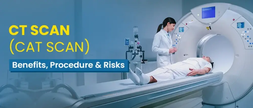
The patient must take the correct position before starting the computed tomography scan. He will be asked to lie on a motorised table that slides into the circular opening of the CT scan machine. The patient must remain still during the scan to ensure clear images for an accurate diagnosis.
The CT scanner has an X-ray tube that rotates around the patient’s body. As it spins, it emits a series of narrow X-ray beams from different angles.
X-ray detectors opposite the X-ray tube capture the X-rays that pass through the patient’s body. Each rotation produces numerous cross-sectional images, or slices, of the area being examined for diagnosis.
After the patient’s scan, a powerful computer processes these X-ray beams and converts them into detailed, cross-sectional images of the body. These slices are then stacked together to form a three-dimensional image, providing a comprehensive view of the patient’s body’s internal structure.
Finally, the radiologist reviews and analyses the scans for abnormalities such as tumours, fractures, infections, or other medical conditions. After the review, they share a detailed report with the patient’s referring doctor.
Not all CT scans are the same; different types focus on various areas of the body, allowing doctors to diagnose conditions more accurately. Let’s take a closer look at the different types of CT scans:
CT scans can be done with or without contrast dye, and each type helps us see different things. Here’s a helpful table that compares these two types of CT scans, making it easier for you to understand the differences.
| Feature | Contrast CT scans | Non-contrast CT scans |
|---|---|---|
| Imaging Detail | Provides enhanced images that highlight blood vessels, organs, and abnormal tissues, aiding in more precise detection of tumours, infections, and inflammation. | Provides basic images of bones and tissues, and is also suitable for identifying structural changes, such as fractures or kidney stones. |
| Contrast Agent | Uses an oral or IV contrast dye to enhance the visibility of organs, blood vessels, and abnormal areas. | No dye is used, making it safer for those with allergies or kidney issues. |
| Preparation | Fasting for a few hours may be needed. Allergy checks ensure the safety of contrast dye. Patients with kidney issues may require additional tests before the scan. | Minimal preparation needed; typically, regular eating and drinking are allowed before the scan. |
| Best For | Ideal for detecting tumours, vascular diseases, inflammation, infections, and other conditions requiring enhanced tissue differentiation. | Ideal for evaluating bones, lungs, trauma, kidney stones, or conditions where detailed soft tissue contrast isn’t crucial. |
| Speed | A longer procedure is required as the contrast dye needs to spread before imaging. | Usually faster as it involves no dye. |
The use and choice between the two depend on the specific medical condition and the patient’s overall health.
It is an advanced imaging technique that combines positron emission tomography (PET) and computed tomography (CT) to provide detailed information about the body’s anatomy and metabolic activities. It is often used to diagnose and manage cancer.
PET-CT scans help detect cancer early by identifying areas of increased metabolic activity. They determine the extent of the tumour and help plan an appropriate treatment. PET-CT scans also help track the efficacy of cancer treatment by comparing visuals over time and identifying cancer recurrence earlier than other methods. PET-CT scans can detect different types of cancer in the body, such as lung, head, neck, breast, thyroid, melanoma, and lymphoma.
CT scans are a highly valuable tool for examining the inside of your body, helping doctors to promptly and precisely identify injuries, infections, and diseases. Here's a list of some reasons and the specific body parts that your doctor might recommend for a CT scan.
CT Scans are usually performed on an outpatient basis. The procedure might depend on the medical condition and the doctor's recommendation. Here’s what you can expect during a CT scan procedure:
Here’s the comparison between CT scans and other types of imaging techniques in India:
| Features | CT Scans | X-ray | Ultrasound | MRI | PET Scan |
|---|---|---|---|---|---|
| Purpose | Detailed cross-sectional images of bones, soft tissues, and blood vessels | Ideally suitable for visualising bones and identifying fractures | Viewing soft tissues, organs, and guiding procedures | Imaging soft tissue, brain, spinal cord, and joints | Detecting cancer, monitoring treatment, responding, and examining brain function |
| How it works | Uses X-rays from multiple angles and computer processing to create detailed images | Uses a small amount of ionising radiation to capture images | Uses high-frequency sound waves to create real-time images | Uses strong magnetic fields and radio waves to generate images | Uses a small amount of radioactive material to show functional processes |
| Strengths | It provides detailed images quickly and is suitable for bone and soft tissue | Quick, widely available, and expensive | No radiation exposure is ideal for soft tissue and fluid-filled structures | Superior soft tissue contrast; no ionising radiation | Shows functional imaging and cellular activity |
| Limitations | Uses ionising radiation, which is relatively expensive | Limited detail, especially for soft tissues | Less detailed and can’t penetrate bone or air-filled spaces well | More expensive, longer scan times are not suitable for patients with metal implants | Lower spatial resolution involves radiation exposure |
| Best used for | Diagnosing complex conditions, emergencies, internal injuries, and cancers | Detecting fractures and bone abnormalities | Monitoring pregnancies, abdominal organs, and blood flow | Soft tissue injuries, brain and spinal cord conditions, and joint issues | Cancer detection, evaluating metabolic and biochemical activity |
| Speed | Moderate (10–30 minutes) | Very fast | Fast (15–45 minutes) | Slow (30–90 minutes) | Moderate (30–60 minutes) |
| Radiation Exposure | Yes (moderate) | Yes (low) | None | None | Yes (low) |
| Cost (Approx.) | Rs. 1,500–Rs. 25,000 | Rs. 300–Rs. 1,000 | Rs. 500–Rs. 2,500 | Rs. 6,000–Rs. 25,000 | Rs. 10,000–Rs. 50,000 |
| Accessibility | Widely available in urban areas | Widely available | Available even in smaller towns | Limited in smaller towns | Very limited in smaller town |
CT scans are a safe and non-invasive way to examine the inside of the body; however, it's essential to be aware of potential side effects.
Minimise the risks and side effects of the CT scan by following these steps:
Before undergoing a CT scan, it’s highly recommended that you consider the following medical conditions:
Inform your healthcare provider about these conditions to help plan the scan safely and effectively.
>> Read More: Health Insurance Costs and Role of Fitness Programs
A CT scan is the most common test for diagnosing underlying medical conditions. It is a vital tool for modern medicine, providing detailed, high-resolution imaging that surpasses regular X-rays. It is versatile and accurately diagnoses most conditions, from tumours to cancers. For any treatment diagnosis, a body scan is essential; if you have health insurance coverage, your expenses can be mitigated. Therefore, choose the best health insurance to protect your financial future.
Disclaimer: The above information is for reference purposes only. Kindly consult your general physician for verified medical advice. The health insurance benefits are subject to policy terms and conditions. Refer to your policy documents for more information.
How to Download Care Health Insurance Policy? Mudit Handa in Health
How to Check the Status of Your Health Insurance Policy? Care Health Insurance in Health
What is the Use of ABHA Health Card? Mudit Handa in Health
How Does a Care Health Insurance Cashless Network Hospital Work Rashmi Rai in Health
Copay vs Consumables: Avoid Confusion in Your Health Insurance Jagriti Chakraborty in Health
Ultimate Care Senior: Best Health Insurance For Senior Citizens! Sejal Singhania in Health
The Power of the Policyholder: What Should You Know? Jagriti Chakraborty in Health
Why Group Medical Insurance is Important for Employees? Riya Lohia in Health
A CT scan produces detailed cross-sectional images of the body to diagnose injuries, infections, tumours, and other conditions, revealing high-resolution images of internal structures such as the brain, chest, abdomen, and spine.
Most CT scans last between 10 and 30 minutes, depending on the body part being imaged and whether contrast dye is administered.
Yes, CT scans are generally safe due to their low radiation levels, and the benefits for diagnosis usually outweigh the minimal risk.
Yes, you can eat and drink normally after the scan unless your doctor has given you different instructions. If contrast dye was used, drinking plenty of water will help eliminate it more quickly.
Most people do not experience any side effects. Rarely, mild allergic reactions such as rash or nausea may occur due to the contrast dye.
A CT scan report describes abnormalities such as masses, lesions, or fractures, including their size, shape, and location. If there are no issues, it states that there are no abnormalities.
Always stay up to date with our newest articles sent direct to your inbox
Loading...